Bibliography for Last Post
Patrick, Ruth. Charles W. Reimer. 1996. The Diatoms of the United States. The Academy of Natural
Sciences of Philadelphia. 19th and the parkway, Philadelphia Pennsylvania 19103. Livingston
Publishing Company. 688.
D.J. Patterson. 1996. Free-Living Freshwater Protozoa: A Colour Guide. 1996. 73 Corringham Road, London NW 117DC UK. Manson Publishing Ltd. 223.
Handbook of Algae, by Herman Silva Frost.
Sunday, November 23, 2014
Last Post
This is the last post I'm going to make here. I just realized exactly what time it is on which day, so it's now. Yes, silly and cuckoo and I really shouldn't have waited this long... Buuuut family stuff interfered. So here it is. And tonight I finish my freaking term lab report to turn in on Tuesday.


1. This is a Fragileria. This guy was new, and it's a pity that the experiment ended so soon. It's a diatom, I've found, and it looks very different from the side. Unfortunately, I couldn't get a picture of the side as it could only be seen from this particular viewpoint.
2. This is a pic of an Anisonema. Honestly, I've seen them before, but I wanted to get a picture that showed both flagella better than the previous one. So, here it is.
3. This picture is of a Pseudomicrothorax. Yes, I know it's a mouthful of a name. I had difficulty spelling it correctly, if I remember the lab right. Anyways, this picture is obviously a little bit blurred. Hopefully you can still tell that this little guy is tinted green, hinting at photosynthetic activity. The blurriness, as usual, is caused when the organism is a bit too fast for the camera to capture properly.
4. This is Cladophora sp., a photosynthetic algae. We found it near the end of the observation, and decided to take a photo despite having a lot of images.
5. The Merisomepedia tranquilla looks a little like a tiny cell city. It's a cluster of algae cells, and there seems to be a carcass floating almost inside the organism.
6. This Nodularia is the very last picture I took. It's an algae that's looks much like a miniature caterpillar.
Here's a further list of what else I found for the last observation of the lab:
Halteria
Nematoads
Tachysoma
Anisonema
Cyclidium
Loxophyllum sp.
Analyds
Vorticella
2 Amoeba
A cluster of freshwater jellyfish
Oscilatoria
Dero Digitata
Well... that's about it. Here the experiment ends, as the MicroAquarium was cleansed on Friday. I'm not sure I did such a great job of this project... But I managed to get posts up for five observations. I'm happy that I managed to finish this, and did not burn out halfway through. I actually quite enjoyed observing the little organisms.
Here the story ends
Of tiny lives and one big friend
Not the traditional tale,
it's true
But a project as big as a whale
To a young Freshman who really hopes that you won't rue
the time you spent reading her tale.
Sunday, November 16, 2014
Bibliography for Almost Last Post
Specific Citations
Anisonema sp. can be found in Free Living Freshwater Protozoa: A Colour Guide on page 54, figure 78.
Dero Digitata can be found on page 301, figure A of Freshwater Invertebrates of the US Protozoa to Mollusca.
Loxophyllum can be found in Free Living Freshwater Protozoa: A Colour Guide on page 132, figure 283-4
The immature Nauplius Cyclops can be found on page 415, figure 6 of Freshwater Invertebrates of the US Protozoa to Mollusca.
Nematoda can be identified on page 230-33 of Guide to Microlife
Heterophrys can be found on page 173, figure 404 of Free Living Freshwater Protozoa: A Colour Guide
General Citations
D.J. Patterson. 1996. Free Living Freshwater Protozoa: A Colour Guide. 1996. 73 Corringham Road,
London NW117D, UK. Manson Publishing Ltd. 223.
Rainis, Kenneth G. Bruce J. Russel. 1996. Guide to Microlife. 1996. Danbury, Connecticut. Franklin
Watts.
Robert W. Pennack. 1989. Freshwater Invertebrates of the Us Protozoa to Mollusca. 1989.
Saturday, November 15, 2014
Almost the Last Post
I have been having some serious spelling issues lately... Hopefully, this post will be free of any possible errors. I've sourced many of these images, and some of my previous ones. The water in the tank had dropped severely when I checked it this time. There were also many strings of cells, as well as several colonies of cells that were difficult to identify as living or dead.


There are also some organisms I spotted, but didn't grab pictures of this time around.
Here's a list:
At least one Nematoda
At least one Paranema
Many Vorticella sp. (they've showed up before!)
Lots of Halteria
One Halteria that looked ready to divide, as the movements were slowed down considerably and it was bigger than it normally was
At least one Heterophrys
2 Difflugia (showed up at least once before, last week)
Analyd that was either poking its head out of its tube, or building the tube it remains in for the rest of its life
Several Loxophyllum (they've showed up before)
There were a lot of worm droppings, just like last week.
My water, as usual, was originally found at:
Water pool below spring. Fountain City Park west of Broadway at Hotel Ave. Knox Co. Knoxville TN. Full shade exposure Spring Feed Pond N36 02.253 W83 55.986 990 ft 10/12/2014
1. This is a Coleps sp. It's very fast, almost too fast for the camera to capture. This is about the best picture I could get of it. It seemed to be dark green in color, and it has a bluish rim around the inner cell that seems to be a membrane from the glimpses I could catch of this speedy organism.
2. I found Dero Digitata in my MicroAquarium this week. This worm was identified by the spike configuration on its back end, pictured above. The Dero seems to like hiding in the bottom layer of the MicroAquarium, poking its head out from the dirt to grab plant matter to consume.
3. I found this Anisonema sp. around the middle of the MicroAquarium. At least, the current middle when I took the pic. It's not a great one, because neither of the flagella are visible... But it's the best the camera could catch. Interesting tidbit, even on the microscope, there didn't seem to be a mouth present on this Anisonema sp.
4. I thought this picture was interesting to include. It's of an absolutely gigantic amoeba, one that there wasn't any positive identification for. This thing could barely fit inside the camera's view, thus the cutoff picture. The amoeba seemed to function through those little streams visible in the picture, nutrients being carried through them.
5. This organism turned out to be an immature Nauplius Cyclops. I thought it was really interesting how it looked like a tiny bug instead of something you'd find in a MicroAquarium climate. Or, well, a spider... Which is a little scary. It moved in an extremely twitchy manner, jerking quickly through the water.
There are also some organisms I spotted, but didn't grab pictures of this time around.
Here's a list:
At least one Nematoda
At least one Paranema
Many Vorticella sp. (they've showed up before!)
Lots of Halteria
One Halteria that looked ready to divide, as the movements were slowed down considerably and it was bigger than it normally was
At least one Heterophrys
2 Difflugia (showed up at least once before, last week)
Analyd that was either poking its head out of its tube, or building the tube it remains in for the rest of its life
Several Loxophyllum (they've showed up before)
There were a lot of worm droppings, just like last week.
My water, as usual, was originally found at:
Water pool below spring. Fountain City Park west of Broadway at Hotel Ave. Knox Co. Knoxville TN. Full shade exposure Spring Feed Pond N36 02.253 W83 55.986 990 ft 10/12/2014
Monday, November 10, 2014
I really, really hate those weeks that are so busy you can't even finish a post. Especially when you have so much new information. I had an exam the same day as I gathered my info for this thing, so I probably shouldn't have put in FOUR HOURS at the lab studying. So, I'm sleep-deprived and late, but this is what SHOULD have been last week's blog post.
Hi! Welcome back to A Botany Student's Studies. Here's a new list of what I've found inside my MicroAquarium after missing about a week.


I got some new water from the spray water bottles Mr. McFarland has up in the lab room. A food pill was added the week I missed observation, the one before this one. Otherwise, I think I've listed the current changes. Hopefully I'll manage to post my next blog completely on time...
Ciao for now.
Bibliography
McFarland, Kenneth [Internet] Botany 111 Fall 2014. [cited ADD DATE]. Available from http://botany1112014.blogspot.com/
University of Nebraska-Lincoln Nematology [Internet] [November 10, 2014] Available from
http://nematode.unl.edu/wormgen.htm
Hi! Welcome back to A Botany Student's Studies. Here's a new list of what I've found inside my MicroAquarium after missing about a week.
1. Vorticella sp. - This little organism reminds me a little bit of a spinning top, except it doesn't move a lot. The flagella that shows on the picture seems to anchor it in place. The top portion that spins identifies it as a vorticella because it makes spinning motions. Two of them were found in my MicroAquarium, and they were identified in the book Free Living Freshwater Protozoa on page 113, figure 233.
2. Unknown Rotifier- This little guy looks like Mr. Nightmare from the last time, the Possible Philodena Rotifier. However, the prongs on one end differentiate the two organisms. Mr. McFarland and I looked up Rotifiers, but neither of us could find which one it really was. It looked similar to one of them, but different tails (that's the spiky end on this one) told them apart. Only one was found in the MicroAquarium.
3. "Real Small Thing" - Okay, I circled what I'm really trying to point out in blue. When it was actually moving, one flagella would turn rather like a jerky clock hand while the other flagella anchored this little organism in place. This one is markedly new to the MicroAquarium, and Mr. McFarland couldn't identify it. Whatever it was, it was extremely small and looks sort of green. It's difficult to tell whether or not something has chlorophyll when it's as small as this little organism. This little thing had difficulty showing up on the highest resolution of the microscope I used, so this is the best picture I could get.
4. Surinella robusta - This organism surprised Mr. McFarland. Apparently it's fairly rare. This Surinella robusta was identified from Diatoms of North America by William C. Vinyard. It looks disturbingly like a chainsaw.... Or that could be my strange imagination. It's movement, if at all, was slow.
5. Nematode sp. - this isn't a new organism. I've seen Nematodes in my Aquarium before, but this is the first time I got a picture. These guys were identified by Ken, and I apparently misspelt the name of this organism on the picture... Forgive me. This organism is multicellular, consisting of "approximately 1,000 somatic cells." (University of Nebraska-Lincoln Nematology). Potentially, hundreds of cells could be associated with reproduction for this little guy (University of Nebraska-Lincoln Nematology). They can range from 0.3 mm to over 8 meters! (University of Nebraska-Lincoln Nematology). One of the nematodes was moving like it was high on a sugar rush- quickly and very jerkily.
I looked this up on Google, admittedly, but I found what I believe is a reliable source. It's cited below as well as in text.
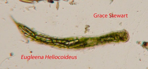
6. Eugleena Heliocoideus- Alright, I misspelt it again. I must have been out of it that particular lab... Anyways. This protozoa has popped up before, and I snagged a picture of it again to show that it had popped up. At least two were spotted, though I only got a picture of one this time. This organism can be identified in a certain algae book I didn't remember (I'll fix it later, I promise), page 283 and figure 386. It doesn't really move.
7. I honestly just thought that this was a cool event to photograph. The amorphous one attempting to engulf the darker one is a chaos amoeba, and the darker one is a shelled amoeba. Eventually, it turned out that the shelled amoeba was too big of a prey for the chaos amoeba, and it moved on. I was simply wowed to witness the chaos amoeba in action... Even if it ended up failing in the end.
I got some new water from the spray water bottles Mr. McFarland has up in the lab room. A food pill was added the week I missed observation, the one before this one. Otherwise, I think I've listed the current changes. Hopefully I'll manage to post my next blog completely on time...
Ciao for now.
Bibliography
McFarland, Kenneth [Internet] Botany 111 Fall 2014. [cited ADD DATE]. Available from http://botany1112014.blogspot.com/
University of Nebraska-Lincoln Nematology [Internet] [November 10, 2014] Available from
http://nematode.unl.edu/wormgen.htm
Thursday, October 23, 2014
Second Posting Day
October 22, 2014
Alright then. Today- rather, tonight- I went to the lab for the first time to the lab where our projects are being kept. It took somewhere around two hours, but I did end up with results. As usual, the water source is as follows: Water pool below spring. Fountain City Park west of Broadway at Hotel Ave. Knox Co. Knoxville TN. Full shade exposure Spring Feed Pond N36 02.253 W83 55.986 990 ft 10/12/2014. I haven't, as of yet, added extra water to my Micro Aquarium. That will hopefully occur next week.
There were quite a bit of carcasses in my Micro Aquarium of many and varied organisms. To think, in only a week... So many organisms lived and died. It's a bit surreal. Even so, I saw a fair few organisms moving in the Micro Aquarium, and I managed to nab a few images. What follows is a list of the organisms I managed to photograph and a bit of information on them. Just a notice, I'll probably be updating this at some point before the actual deadline with some information from the Web.

1. This one is an Urocentrum. This little guy likes to spin around, making it look like a toy top. Ken McFarland helped me identify this one, who we found in Free Living Freshwater Protozoa: A Colour Guide by D.J. Patterson on page 184. I've seen at least three of these organisms, but I'm not quite sure if I'm counting correctly due to the fact these guys move FAST.

2. Litonotous Cygnus was found inside my Micro Aquarium as well. This skinny fellow was identified in the same book as the Urocentrum, Free Living Freshwater Protozoa: A Colour Guide by D.J. Patterson. However, this one was on page 132. It moved very swiftly and fluidly, and was difficult to snap a clear photo of. I spotted two L. Cygnus, at least.

3. This one looked like something out of my nightmares. The organism wasn't positively identified, but Mr. McFarland theorizes that it could be a Philodina Rotifier, in some early stage or constricted by the environment of the Micro Aquarium. It actually looks somewhat similar to the aforementioned organism, but it did not once reveal the rotating sections that characterize a Rotifier during observation. I'll be interested to attempt finding this one again... If I can get over the fact that this organism looks like a tiny dinosaur. This guy moved around fairly actively, seemingly moving by himself. This is the only one that I spotted.

4. This is an actual Philodena Rotifier, more specifically a Philodena sp. Above are shown when the "rotifers" are out, and when they are retracted as a comparison of the two. This was the only one of it's type (confirmed, at least) that I spotted. It was identified by Mr. McFarland. The "rotifers" moved a bit like a helicopter blade might when flattened, but the actual organism didn't do much moving.
5. I was honestly focusing on another organism when Mr. McFarland took a look through the camera screen and spotted this. Apparently, it's a type of algae. It was found in the Handbook of Algae on page 283, written by Herman Silva Frost. I'm glad I got a plant in there, as this class is technically supposed to be about them... I wonder if more will grow in my Micro Aquarium by next week. That would be intriguing, as this was the only one I spotted specifically.
Anyways... That was about the extent of my first visit to Lab Room 507. I'm hoping to find different organisms as well as changes to the ones currently next week. So far, though, this is shaping up to be a very intriguing experiment.
Tuesday, October 14, 2014
First Day of Project
First Day of The Project
Tuesday October 14, 2014
By: Ravenna Nightgale
Alright, today we made our micro aquarium. It was pretty intriguing to make, after we'd gotten done with the mandatory test over last section's materials and the activity for the main part of Lab Class. I'll go over the basics of how the Micro Aquarium was made today, as well as some notes on where the water came from and that kind of thing. Also, there were at least two organisms Mr. MacFarland and I spotted already in my Micro Aquarium.
- We got the Micro Aquarium itself, the base to hold it up, the lid to the Micro Aquarium and sticky to seal the water in.
- We then stuck stickers on the Micro Aquarium itself so we could keep straight whose Aquarium was whose. At least, that was the idea. The pattern was stickers for your section, your table number, and your respective usual seat. My code is green-yellow-red, respectively.
- We were also asked to initial at least two of the stickers as further precaution.
- The students in class divided up to get water from the various sources and dripped it into the Micro Aquariums, along with some of the dirt that had come along with the water.
- After you had gotten both mud and dirt from one of the 12 sources, you would find which two water-weeds you wanted to use in the Aquarium to oxygenate the water (we had to be careful not to obscure the glass, and still get enough to work properly)
- When you finished using the dissection tweezers to put the weed into the Micro Aquarium, you would stick the lid back onto the top using the BlueStick sticky substance given to you earlier.
- Turn it into the box that would take it up to the Lab we would visit to see it again.
I chose water from Source 12, water from a pool below a spring in Fountain City Park with the accompanying soil. Here's the information from the last page on where exactly that is.
Water pool below spring. Fountain City Park west of Broadway at Hotel Ave. Knox Co. Knoxville TN. Full shade exposure Spring Feed Pond N36 02.253 W83 55.986 990 ft 10/12/2014
Water pool below spring. Fountain City Park west of Broadway at Hotel Ave. Knox Co. Knoxville TN. Full shade exposure Spring Feed Pond N36 02.253 W83 55.986 990 ft 10/12/2014
Of course, I added it to my experiment on the 14th of October. Now for the information on the plants used in the Micro Aquarium. I added both of these.
Amblestegium varium (Hedwig) Lindberg. Moss.
Collection from: Natural spring. at Carters Mill Park, Carter Mill Road, Knox Co. TN. Partial shade exposure. N36 01.168 W83 42.832. 10/12/2014
Utricularia gibba L. Flowering plant. A
carnivorous plant. Original material from south shore of Spain Lake (N 35o55 12.35" W088o20' 47.00), Camp Bella Air Rd. East of Sparta Tn. in White Co. and grown in water tanks outside of greenhouse at Hesler Biology Building. The University of Tennessee. Knox Co. Knoxville TN.
10/12/2014
Since this is the first day these things were put together, there wasn't much to be observed in my Micro Aquarium. Mr. MacFarland did spot a seed shrimp, and I spotted what might be a nimitoad. Mr. MacFarland identified both of them. I also didn't get any images, as we haven't gotten onto the microscopes that can do that kind of thing yet. It was really intriguing watching the seed shrimp burrowing into the soil, though... I'd never seen something like it before.
Subscribe to:
Posts (Atom)